
MRI or CT — which to choose? CT is better for bones, lungs and emergencies, while MRI is better for the brain, joints and soft tissue. We explain the difference between the methods, their safety, contraindications, preparation and how the scan is done.
What is the difference between CT and MRI
The two methods rely on different physics. Computed tomography (CT) uses X-rays: the scanner takes a series of images from different angles and a computer builds cross-sections from them. Magnetic resonance imaging (MRI) uses no radiation — it relies on a strong magnetic field and radio waves acting on hydrogen atoms in the tissues.
Which is better: CT or MRI
Neither method is “better” overall — they answer different questions.
CT is chosen to assess:
- the lungs — pneumonia, nodules, lung cancer;
- bones and complex fractures;
- emergencies — trauma, bleeding in the brain, suspected stroke;
- kidney and ureteric stones;
- blood vessels (CT angiography), including the coronary arteries;
- tumour spread in the chest and abdomen.
MRI is more informative for:
- the brain and spinal cord — see brain MRI;
- the spine and intervertebral discs — spine MRI;
- joints, ligaments and menisci — joint MRI;
- pelvic organs, the liver and the breasts;
- soft tissue, when the difference between healthy and abnormal tissue is small.
In oncology the methods often complement each other, and PET-CT is used to look for metastases throughout the body.
Which method is safer
MRI involves no radiation, so it can be repeated as often as needed and used in children. CT involves X-ray exposure; modern scanners use low-dose protocols, and the benefit of a justified scan outweighs the risk. There is no strict “once a year” limit: the doctor decides how often CT is needed, and it should not be done “just in case”.
Contraindications
CT: pregnancy (the scan is done only for vital indications). For CT with iodinated contrast — contrast allergy, severe kidney failure, uncontrolled thyroid disease; caution is needed in diabetes treated with metformin and in multiple myeloma.
MRI: pacemakers and implants that are not MRI-compatible, ferromagnetic vascular clips, metal fragments in the eye, some cochlear implants. Modern joint replacements, braces and most stents usually do not interfere, but the staff must be told about them. Open scanners or sedation help with claustrophobia.
Types of scanners
MRI scanners differ in magnetic field strength. 1.5 Tesla is enough for most tasks, while 3 Tesla machines give the most detailed images — important for the brain, prostate and small joints. CT scanners differ in the number of slices — 16, 64, 128 and more: the more slices, the faster the scan and the sharper the images of moving organs such as the heart.
Why contrast is used
A contrast agent highlights blood vessels and areas with increased blood supply, including tumours and inflammation that might otherwise be missed. CT uses iodine-based agents, contrast-enhanced MRI uses gadolinium-based agents. Both are cleared by the kidneys, so a creatinine blood test is done before the scan.
How to prepare
- Special preparation is usually not needed.
- Do not eat for 2–4 hours before a contrast scan.
- Remove jewellery, watches and clothing with metal parts.
- Bring previous images and reports — the doctor needs to compare changes over time.
- Tell the staff about pregnancy, allergies, kidney disease and implants.
How the scan is done
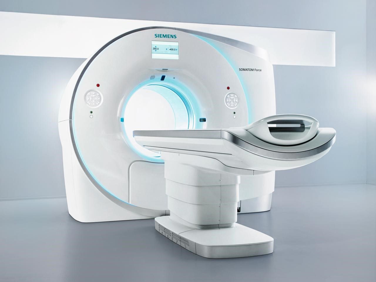
CT takes a few minutes. The patient lies on a table that moves through the ring of the scanner and follows the operator's instructions, such as holding the breath. During contrast injection a feeling of warmth and a metallic taste are possible — this is normal and passes quickly.
MRI takes 15–45 minutes depending on the area. You need to lie still throughout. The scanner makes loud knocking sounds, so the patient is given headphones and a call button to stop the scan if they feel unwell.
Who reports the result and when
The scanner only produces images — a radiologist interprets them. The report is usually ready the same or the next day; complex cases are discussed by a team. The patient receives the report and the images on a disc or via a link. Keep the images themselves, not just the report: they will be needed for a second opinion.
Is a second opinion on the images worthwhile
When a tumour is suspected or before complex surgery, a review of the images by an experienced radiologist at a specialised centre sometimes changes both the diagnosis and the treatment plan. Clinics Direct patients often request exactly this kind of second opinion from doctors in Germany, Israel, Turkey and Spain.
Not sure which scan you need, or want a second opinion on your images? Send a request — Clinics Direct coordinators will suggest a clinic and the cost of diagnostics, free of charge.
